
What are veterinary CT/CAT Scans?
The Regional Veterinary Referral Center is pleased to announce that we offer outpatient CT Scans for your clients. This service will be available by appointment.
Request forms can be found on our website along with information regarding all requirements needed in order for us to perform the CT scan. All procedures are supervised by a licensed veterinarian and the images are read out by a Board-Certified Radiologist.
For additional information regarding this service please contact our Medical Oncology Department at 703-451-8900.
CT Scan Services at The Regional Veterinary Referral Center
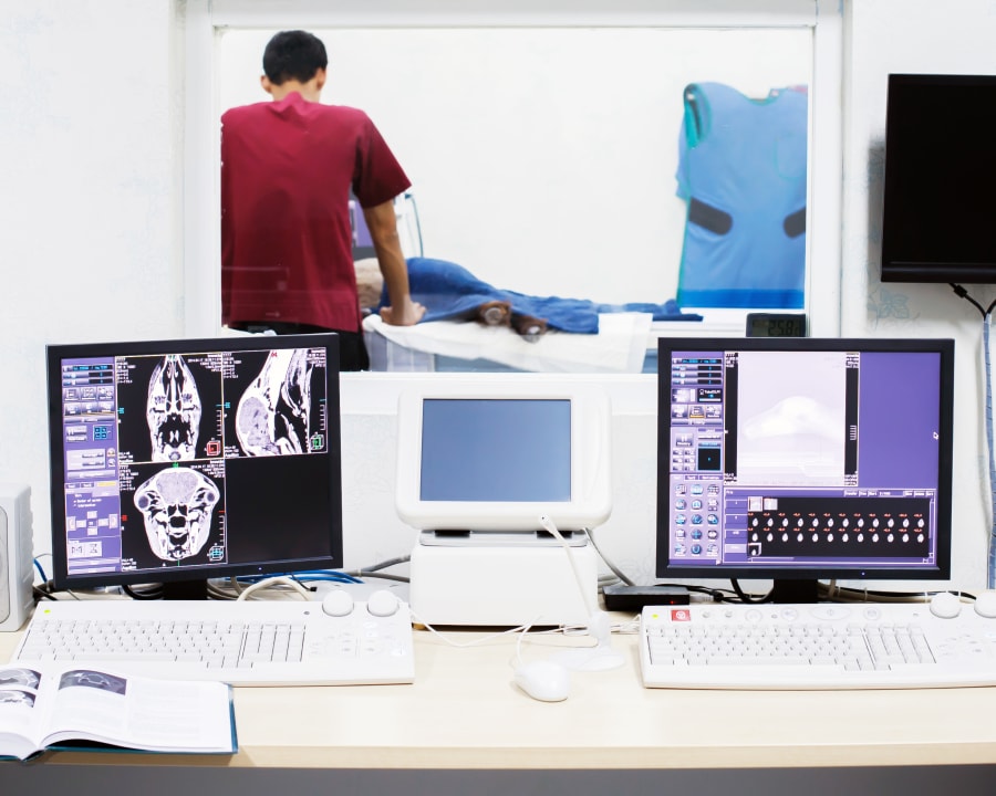
We now have a helical multi-slice scanner. This advanced technology allows us to perform much quicker examinations (reduced time under anesthesia) and gives us even greater detail than was possible before.
Equally as exciting is the addition of software that allows us to convert two-dimensional images into three-dimensional projections! The advantages to us, our referring veterinarians and our patients are innumerable! We can now localize lesions in three dimensions!
This provides better diagnostic information and allows for far more accurate treatment planning. In addition, we can monitor the patients’ response to therapy and alter our treatment plans accordingly to provide the greatest chance of success.
In summary, computed tomography is a safe, beneficial, and advanced imaging technique that will often allow for a more precise and complete diagnosis of a pet’s injury or illness. The information provided by CT – when interpreted by an appropriately trained and skilled veterinary specialist – allows for informed decisions to be made about a pet’s treatment by its owner, general veterinarian, and specialty veterinary team.
All CT images are read by a board-certified Veterinary Radiologist.
What is the difference between CT Scans and X-rays?
CT Scans provide far more detail than traditional X-rays and are important tools in surgical planning for liver tumors, pulmonary tumors, cancers and other conditions. They can tell us the precise location of the tumor and help minimize surgery time.CT Scan Service FAQs
The following are some of the most common questions our veterinary specialists receive about diagnostic procedures involving CAT scans.
- What are the advantages of CT Scans over X-rays?
The computer is able to collect a great deal more information than what is available on a simple X-ray study (radiograph).
In addition, collecting this information for multiple planes allows for a three-dimensional interpretation of the body structure in question. Even more minute detail can then be obtained through computer manipulation of the gray-scale making up the images. This results in images with superior soft tissue differentiation.
Conventional X-rays require a density difference between tissues of at least 10 percent – whereas CT requires a density difference of 0.5 percent or less – before differentiation is detected on the final image. Multiple conventional x-ray exposures of the same body part are often required to evaluate bone and soft tissues separately so that both can be interpreted at their optimal image contrast and density levels.
With CT, the gray-scale can be manipulated so that the bone and each of the soft tissues can be evaluated individually at their optimal contrast and density levels without having to repeat the study.
- What is involved in a CT study?
Because CT requires many x-ray beams to penetrate the body at various angles to create an individual image slice, it is crucial that the patient not move during the procedure. The entire CT imaging process also takes slightly more time than a standard x-ray procedure.
However, due to our new helical multi-slice scanner, we can take as many as 60 high-resolution scans per minute! Anesthesia, and limited pre-anesthesia blood work depending on the individual pet’s medical condition, is therefore necessary.
Your pet will need to be well fasted (an 8-12 hour fast is usually recommended) before anesthesia. A catheter will be placed in your pet’s leg for the anesthesia. This catheter will be used for the administration of anesthetic drugs, fluid therapy and occasionally for contrast agents needed for the study. A technician will be dedicated to monitoring your pet while your pet is under anesthesia.
A variety of equipment will be used for monitoring heart rate/rhythm, breathing rate, levels of oxygen and carbon dioxide, blood pressure and continuous temperature rate. Your pet will be precisely positioned for the study required and may need repositioning if more than one region is being studied. Occasionally, even with the improved image resolution offered by CT, some abnormalities remain undetectable without the use of contrast media – often referred to as a “dye study.” A “dye study” is almost always performed for CT’s of the brain.
The length of time your pet will be under anesthesia will vary depending on the extent of the study. Due to our purchase of a helical multi-slice scanner, we have dramatically reduced scan times and the duration of anesthesia needed to safely perform the scan. For most studies, your pet is likely to be under anesthesia for less than 20 minutes!
Following completion of the study, your pet is allowed to recover from anesthesia while the computer manipulates the acquired data. Once your pet has fully recovered, and is once again completely alert, he/she will be free to go home.
- When could my pet benefit from a CT scan versus conventional X-rays?
CT is still the most advanced imaging modality for evaluating detail of boney structures. Its impressive sensitivity in the detection of subtle bone detail has allowed:
- Detection and evaluation of the extent of bone loss and/or bone repair of subtle stress fractures that are not detectable in conventional x-ray images.
- The evaluation of the extent of bone invasion by primary bone tumors.
- Detection of subtle early bone invasion by adjacent soft tissue tumors.
Due to their limitation in tissue density differentiation, conventional x-rays usually do not provide any information on the internal anatomy of individual organs (except lungs). CT is capable of revealing more information regarding the internal structure of soft tissue organs. Examples of this include:
- Identification of tumor spread within organs (including liver, spleen, kidneys, etc.)
- Internal detail of the spine or brain tissue.
- Differentiation between the soft tissue structures in the chest located in front of the heart (mediastinum) or adjacent to the heart.
- Differentiation between soft tissue structures in the chest when the presence of fluid obstructs their visualization on conventional x-rays.
- Detection of subtle tumor spread within the lung tissue.
- CT has been shown to be eight times more sensitive than conventional x-rays at detecting tumor spread to the lungs!
A conventional x-ray provides a two-dimensional image of a three-dimensional object. Therefore, many structures seen on x-rays are superimposed over one another. This can make it very difficult to evaluate an image, and impossible to precisely interpret it.
Although at least two perpendicular views, on x-rays, are usually taken to achieve a better three-dimensional impression, the acquired information remains limited. CT offers a more accurate three-dimensional impression, which is particularly useful in evaluating complex areas such as the skull, spine and joints.
Examples may include:
- Diagnosis of lumbosacral disease.
- Evaluation of joint surfaces affected by arthritis, infection, trauma or tumor invasion. CT is particularly helpful in the diagnosis of elbow dysplasia.
- CT is unarguably the best way to evaluate the sinuses, nasal and oral cavities for the detection of masses, old or recent fractures or foreign bodies imbedded in these cavities.
- General imaging of the head for the detection of any skull fracture caused by head trauma, masses occurring behind or surrounding the eyes, and abnormalities involving the middle ear and its surroundings.